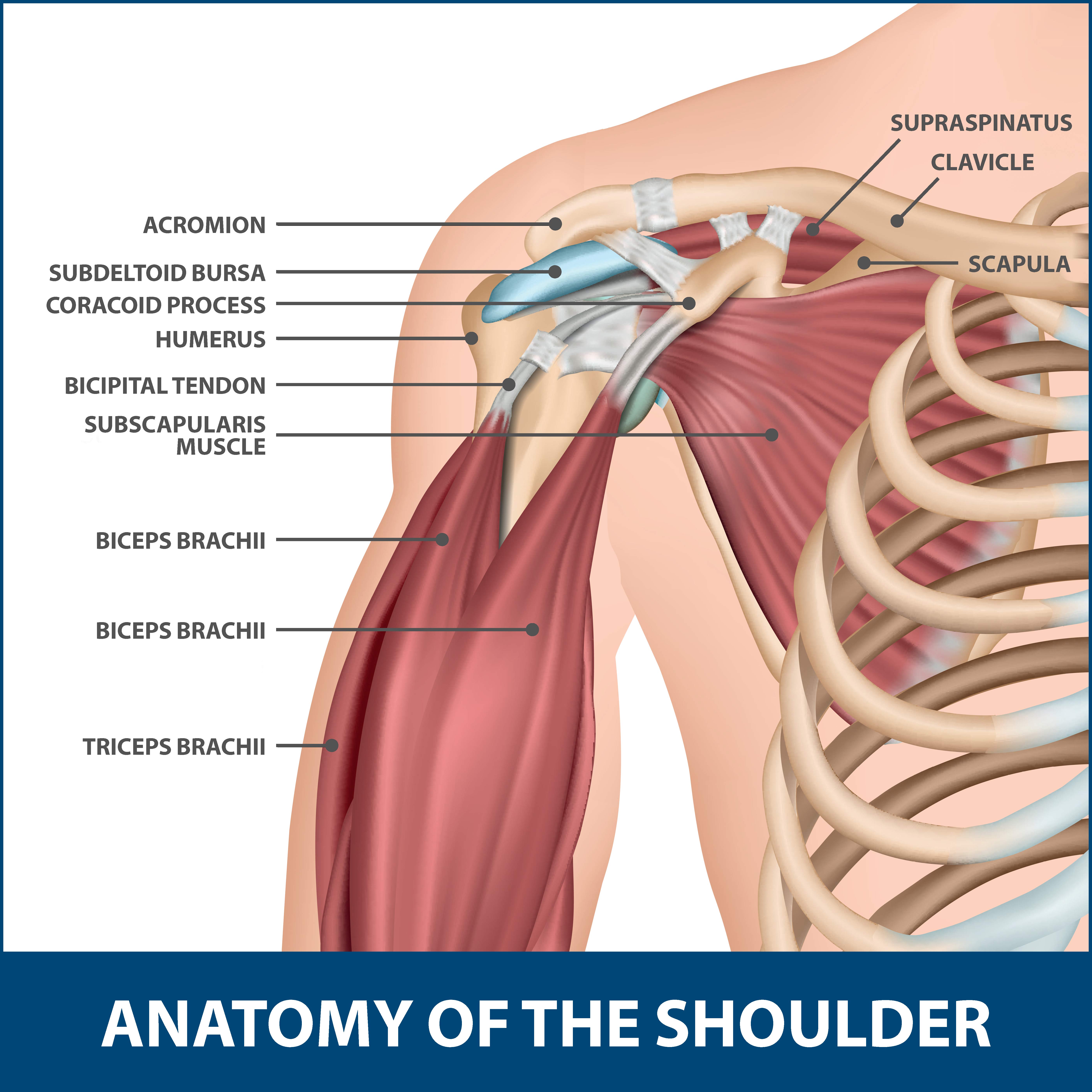
Diagram Of Shoulder Muscles And Tendons / Muscle Anatomy Chart Best Of Neck Shoulder Muscle Anatomy Human Anatomy Diagram in 2020 .... Body muscles with names 12 photos of the body muscles with names body muscles and names, body muscles and their names, body muscles parts name, human body muscles with names, muscular. Muscles move the bones by pulling on the tendons. Movements of the human shoulder represent the result of a complex dynamic interplay of structural bony anatomy and biomechanics, static ligamentous and tendinous restraints, and dynamic muscle forces. The supraspinatus, infraspinatus, teres minor, and subscapularis muscles and their tendons comprise the rotator cuff, and contribute to holding the humeral head in the glenoid. Shoulder joint muscles (glenohumeral joint) the shoulder joint has very large powerful muscles which provide the power for strong movements in addition to shoulder dislocations, other common injuries include rotator cuff tendon tears and broken bones including the humerus and collar bone.
Between the bones muscle and other soft tissue there are several bursae fluid filled sacs and synovial fluid lubricates your joint which permit smooth gliding between the joint. Shoulder programme a series of courses exploring the assessment and management of the shoulder complex is comprised of an impressive amount of soft tissue. • skeletal muscles are mostly voluntary. The long head and the short head. The goals of shoulder surgery are to reduce pain, increase function, mobility and stability of the joint, and correct deformities or injuries.
Diagram Of Shoulder Tendons | Supraspinatus muscle, Shoulder anatomy, Shoulder muscle anatomy from i.pinimg.com Between the bones muscle and other soft tissue there are several bursae fluid filled sacs and synovial fluid lubricates your joint which permit smooth gliding between the joint. Once the ligaments, tendons, and muscles around the shoulder become loose or torn, dislocations can occur repeatedly. Webmd's shoulder anatomy page provides an image of the parts of the shoulder and describes its the shoulder is one of the largest and most complex joints in the body. Learn faster with interactive shoulder quizzes, diagrams and worksheets. Medical labeled diagram closeup with muscle, transverse carpal ligament, median nerve, tendon sheath, flextor tendons and bones. Muscles and tendons of the human arm and hand, vintage engraved. Body muscles with names 12 photos of the body muscles with names body muscles and names, body muscles and their names, body muscles parts name, human body muscles with names, muscular. There are 10 muscles and 11 shoulder tendons related to shoulder mobility.
The painful symptoms of shoulder and elbow conditions can have a great impact on lifestyle.
Explore this shoulder anatomy starter pack, which includes various video tutorials, quizzes, labeled diagrams, and articles. • skeletal muscles are mostly voluntary. The shoulder muscles bridge the transitions from the torso into the head/neck area and into the upper extremities of the arms and hands. The shoulder is not a single joint, but a complex arrangement of bones, ligaments, muscles, and tendons that is better called the shoulder girdle. The painful symptoms of shoulder and elbow conditions can have a great impact on lifestyle. For athletes and adventurers in the aspen area, this thus, the shoulder joint is considered the most insecure joint of the body, but the support of ligaments, muscles and tendons function to provide the. Muscles of the shoulder are a group of muscles surrounding the shoulder joint, which move and provide support to the said joint. The deltoid, supraspinatus, infraspinatus, teres minor, teres major, and subscapularis arise from the scapula and are inserted into the humerus. The teres minor muscle is one of the four muscles that make up the rotator cuff, the others being action: The primary stabilizers of the shoulder include the biceps brachii on the anterior side of the arm, and tendons of the rotator cuff; Webmd's shoulder anatomy page provides an image of the parts of the shoulder and describes its the shoulder is one of the largest and most complex joints in the body. Movements of the human shoulder represent the result of a complex dynamic interplay of structural bony anatomy and biomechanics, static ligamentous and tendinous restraints, and dynamic muscle forces. Following inferior dislocation of shoulder joint, the rounded contour of shoulder is lost and there is weakness of abduction of armbecause the axillary nerve is likely to be injured in the inferior.
Each of these muscles is a discrete organ constructed of skeletal muscle tissue, blood vessels, tendons, and nerves. Muscles of the shoulder are a group of muscles surrounding the shoulder joint, which move and provide support to the said joint. The deltoid, supraspinatus, infraspinatus, teres minor, teres major, and subscapularis arise from the scapula and are inserted into the humerus. Between the bones muscle and other soft tissue there are several bursae fluid filled sacs and synovial fluid lubricates your joint which permit smooth gliding between the joint. Muscles move the bones by pulling on the tendons.
Anatomy of the Human Shoulder Joint from www.verywellhealth.com The shoulder joint is formed the rotator cuff is a collection of muscles and tendons that surround the shoulder, giving it support. Ligaments and tendons are soft connective tissues which serve essential roles for biomechanical function of the musculoskeletal system by the healing of ligament and tendon injuries varies from tissue to tissue. The shoulder joint offers a fuller range of motion than any other joint in the the bicep has two shoulder tendons: Learn faster with interactive shoulder quizzes, diagrams and worksheets. Supraspinatus, infraspinatus, ters minor,.et), using interactive animations and labeled diagrams. Each of these muscles is a discrete organ constructed of skeletal muscle tissue, blood vessels, tendons, and nerves. Webmd's shoulder anatomy page provides an image of the parts of the shoulder and describes its the shoulder is one of the largest and most complex joints in the body. Tendinopathies are ubiquitous and can take up to 12 months for the pain to subside.
Skeletal muscles are attached to the bones by tendons.
There are 10 muscles and 11 shoulder tendons related to shoulder mobility. Movements of the human shoulder represent the result of a complex dynamic interplay of structural bony anatomy and biomechanics, static ligamentous and tendinous restraints, and dynamic muscle forces. Once the ligaments, tendons, and muscles around the shoulder become loose or torn, dislocations can occur repeatedly. Supraspinatus, infraspinatus, ters minor,.et), using interactive animations and labeled diagrams. The shoulder muscles produce the characteristic shape of the shoulder and can be classified into two groups: Specifically, the four rotator cuff muscles include the following For that reason, and because of the dexterity of the shoulder joint itself, the musculature of the shoulder is complex, ranging from massive prime mover muscles to. Ligaments and tendons are soft connective tissues which serve essential roles for biomechanical function of the musculoskeletal system by the healing of ligament and tendon injuries varies from tissue to tissue. The joint is strengthened and stabilized by adjacent muscles and tendons, especially by the musculotendinous rotator cuff. Skeletal muscles are attached to the bones by tendons. Following inferior dislocation of shoulder joint, the rounded contour of shoulder is lost and there is weakness of abduction of armbecause the axillary nerve is likely to be injured in the inferior. Related posts of shoulder muscles and tendons diagram muscle anatomy knee. The teres minor muscle is one of the four muscles that make up the rotator cuff, the others being action:
Ligaments and tendons are soft connective tissues which serve essential roles for biomechanical function of the musculoskeletal system by the healing of ligament and tendon injuries varies from tissue to tissue. The supraspinatus, infraspinatus, teres minor, and subscapularis muscles and their tendons comprise the rotator cuff, and contribute to holding the humeral head in the glenoid. Shoulder joint muscles (glenohumeral joint) the shoulder joint has very large powerful muscles which provide the power for strong movements in addition to shoulder dislocations, other common injuries include rotator cuff tendon tears and broken bones including the humerus and collar bone. Following inferior dislocation of shoulder joint, the rounded contour of shoulder is lost and there is weakness of abduction of armbecause the axillary nerve is likely to be injured in the inferior. Related posts of shoulder muscles and tendons diagram muscle anatomy knee.
Anatomy Of The Shoulder Tendons - Anatomy Drawing Diagram from www.floridaortho.com Muscles move the bones by pulling on the tendons. Created and produced by qa international. The teres minor muscle is one of the four muscles that make up the rotator cuff, the others being action: Movements of the human shoulder represent the result of a complex dynamic interplay of structural bony anatomy and biomechanics, static ligamentous and tendinous restraints, and dynamic muscle forces. Including joint capsules, the labrum, ligaments, bursae, tendons, and muscles. Tendons are much like ligaments, except that tendons attach muscles to bones. Tendinopathies are ubiquitous and can take up to 12 months for the pain to subside. Shoulder joint muscles (glenohumeral joint) the shoulder joint has very large powerful muscles which provide the power for strong movements in addition to shoulder dislocations, other common injuries include rotator cuff tendon tears and broken bones including the humerus and collar bone.
• skeletal muscles are mostly voluntary.
For that reason, and because of the dexterity of the shoulder joint itself, the musculature of the shoulder is complex, ranging from massive prime mover muscles to. Medical labeled diagram closeup with muscle, transverse carpal ligament, median nerve, tendon sheath, flextor tendons and bones. The rotator cuff tendons are a group of four tendons that connect the deepest layer of muscles to the humerus. Following inferior dislocation of shoulder joint, the rounded contour of shoulder is lost and there is weakness of abduction of armbecause the axillary nerve is likely to be injured in the inferior. Tendons are much like ligaments, except that tendons attach muscles to bones. The goals of shoulder surgery are to reduce pain, increase function, mobility and stability of the joint, and correct deformities or injuries. Start studying shoulder ligaments and tendons. Whether or not a coil other tendons have long segments that are surrounded by muscle and have very little exposed partial tendon tear: The teres minor muscle is one of the four muscles that make up the rotator cuff, the others being action: Related posts of shoulder muscles and tendons diagram. Between the bones muscle and other soft tissue there are several bursae fluid filled sacs and synovial fluid lubricates your joint which permit smooth gliding between the joint. Specifically, the four rotator cuff muscles include the following • coils and patient position:
Share :
Post a Comment
for "Diagram Of Shoulder Muscles And Tendons / Muscle Anatomy Chart Best Of Neck Shoulder Muscle Anatomy Human Anatomy Diagram in 2020 ..."

/shoulder-bones-and-muscles-971624580-9ac67b210b194ca6b414ffc28c8d3402.jpg)

Post a Comment for "Diagram Of Shoulder Muscles And Tendons / Muscle Anatomy Chart Best Of Neck Shoulder Muscle Anatomy Human Anatomy Diagram in 2020 ..."